A whole world that we never see exists under the lens of a microscope. For the past 11 years, talented people have lived microscopic life on film for the Nikon Small World in Motion Microphotography Competition. The winning entries brought a tiny world into focus by recording video footage or digital time-lapse footage using a microscope. The entries were then judged by a jury of experts from the fields of microphotography and photography.
This international competition has been creatively combining science and technology since 1975. In 2011, Nikon added a video category to its microphotography competition called Small World in Motion. Since then, the five best films that skillfully stage the little world under the lens were awarded places one to five every year.
Here are the top five winning entries of 2021, capturing a tiny world in motion:
#1. Microfauna in a termite belly
(Courtesy Nikon Small World / Fabian J. Weston)
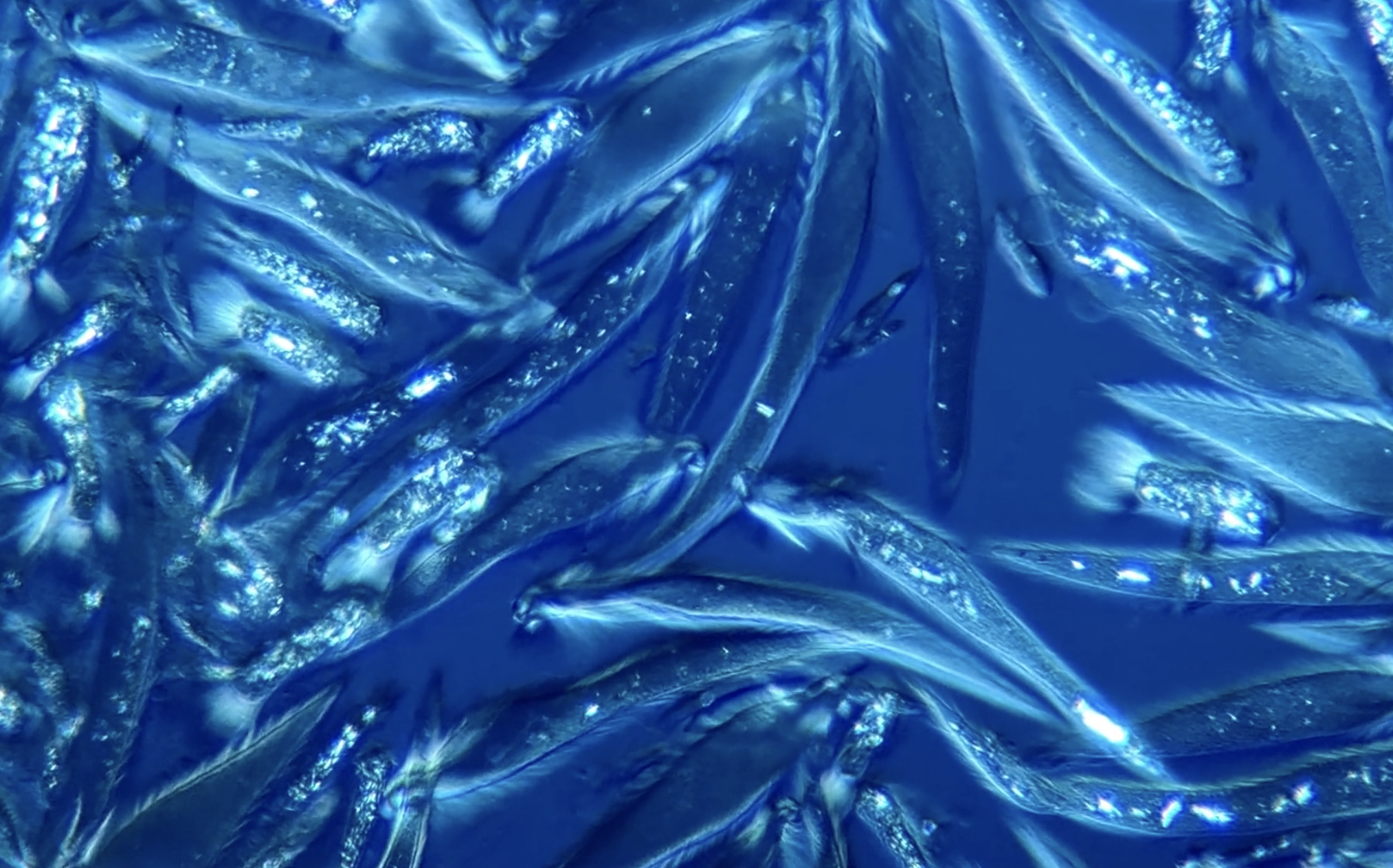
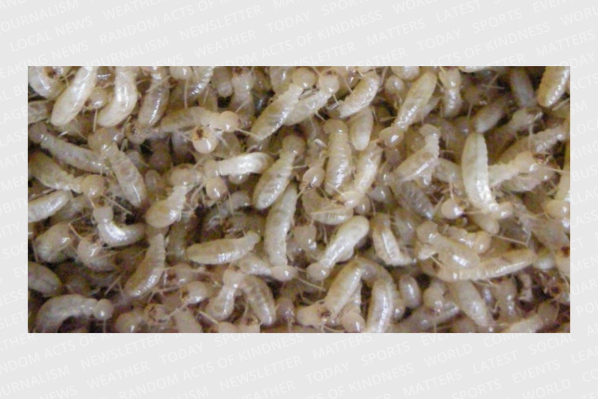
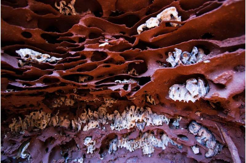
Fabian J. Weston of Pennant Hills, New South Wales, Australia won first place for capturing microscopic animals known as microfauna in the belly of a termite. In his video, viewed at 10X, 20X, and 40X, you can see that these tiny creatures play an important role in feeding the termites. They help digest cellulose, the main food for termites.
# 2. A human microtumor forms and spreads
(Courtesy Nikon Small World / Stephanie Hachey and Christopher Hughes)
In this 10-day time-lapse that shows an artificial microtumor forming and metastasizing, University of California molecular biologists Stephanie Hachey and Christopher Hughes created a special environment in their laboratory that has been controlled for CO2 and moisture. The scientists used confocal and Fluorescence imaging in their work a technique that enabled them to clearly illuminate a seldom seen biological process. Here, at 10x magnification, blood vessels (in red) support the growing tumor (in blue).
# 3. A water flea mom gives birth to little boys
(Courtesy Nikon Small World / Andrei Savitsky)
Observe several tiny water fleas (Daphnia pulex) swim away just seconds after their mother is born. Andrei Savitsky from Cherkassy, Ukraine captured this amazing video at 4x magnification. A technique called Dark field microscopy illuminated these tiny aquatic creatures and made them visible in their natural habitat.
# 4. Nerve fibers in motion after crossing the midline of the central nervous system
(Courtesy Nikon Small World / Alexandre Dumoulin)
Immerse yourself in the central nervous system and observe commissural axons (Nerve fibers) in motion as they cross the midline, the organizing center of the nervous system. Alexandre Dumoulin from the University of Zurich confocal imaging at 40X magnification to highlight these nerve fibers that convey information and connect the two sides of bilateral animals.
# 5. An infected mosquito chases malaria parasites
(Courtesy Nikon Small World / Sachie Kanatani and Photini Sinnis)
See Johns Hopkins molecular biologists Sachie Kanatani and Photini Sinnis catch an infected mosquito that is salivating on malaria parasites. They used confocal imaging at 10x magnification to clearly focus this action.


