Japanese camera and optical equipment maker Nikon has announced the winners of its 11th annual Small World in Motion competition, which highlights some of the best current photography and video footage under the microscope.
The winning pictures and video clips give a breathtaking insight into a microscopic world that normally remains hidden from us.
The first prize of this year’s competition went to Fabian Weston from Pennant Hills, New South Wales, Australia, who took fascinating pictures of microorganisms in the intestines of a termite.
Weston recorded the video using a research microscope from the 1970s. Its aim was to visually illustrate the symbiotic relationship between termites and these particular microscopic organisms.
The organisms and their termite host were difficult to film because they are sensitive to light and oxygen. Even small changes in the environment can lead to the death of termites and their symbiotic microorganisms.
“The biggest challenge in making this video was figuring out the right solution for the creatures themselves,” Weston said in a statement. “I’ve tried many methods, even making my own saline solution.”
“They are very sensitive to oxygen, so I had to remove as much gas from the solution as possible. It was very tricky and I had to work quickly. The video you see is the result of months of trial and error, a lot of research and perseverance. “
The microorganisms observed in the termite intestine are known as protists – a term that refers to a diverse group of predominantly unicellular organisms that are not considered to be plants, animals, or fungi.
The organisms seen in the video have developed a unique relationship with termites by digesting the plant cellulose that the insects eat and helping them make food from it, while at the same time returning carbon to the soil.
Tumor and a water flea
Second place in the competition went to Dr. Stephanie Hachey and Dr. Christopher Hughes of the Department of Molecular Biology and Biochemistry at the University of California, Irvine, for her fascinating time-lapse of artificial tumor formation and spread.
Other winners included Andrei Savitsky from Cherkassy, Ukraine, who took third place, who took pictures of a water flea (Daphnia pulex) giving birth to tiny boys.
In the meantime, Dr. Sachie Kanatani and Dr. Photini Sinnis of the Department of Molecular Microbiology and Immunology at the Johns Hopkins Bloomberg School of Public Health, who recorded an infected mosquito feeding malaria parasites that had been tagged with a fluorescent substance.
The honorable mentions included Sophie-Marie Aicher and Dr. Delphine Planas from the Department of Virology at the Institut Pasteur Paris in France, who recorded images of infection with SARS-CoV-2 – the virus that causes COVID-19 – which triggers cell fusion and cell death in bat brain cells.
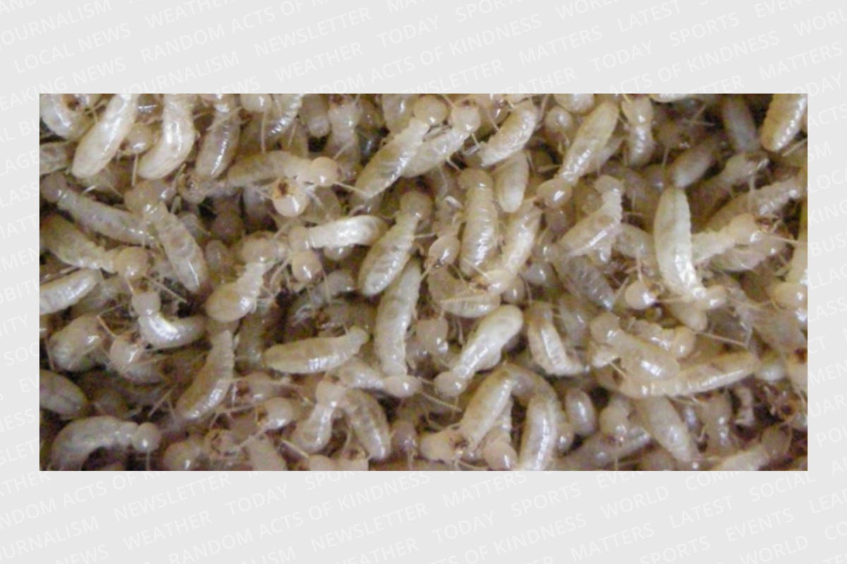
This screenshot is from a video showing microorganisms in the intestines of a termite. It won first prize in the 2021 Nikon Small World In Motion Microphotography Contest.
Nikon Small World In Motion / Fabian J. Weston


